Horse Leg Bone Diagram - Disorders Of The Shoulder And Elbow In Horses Horse Owners Veterinary Manual / Cannon bone this is also called the 3rd metacarpal and has the splint bones running down either side of it;
Horse Leg Bone Diagram - Disorders Of The Shoulder And Elbow In Horses Horse Owners Veterinary Manual / Cannon bone this is also called the 3rd metacarpal and has the splint bones running down either side of it;. For more anatomy content please follow us and visit our website: License image the bones of the leg are the femur, tibia, fibula and patella. The photograph shows the laminae which keep the hoof wall tightly bonded to the internal structures. It is made up of the ilium, the ischium, and the pubis.at the junction of these three bones is a cavity called the acetabulum, which acts as the socket of the hip joint.the pelvic cavity is larger in diameter in the mare than in the stallion, providing more room for the foal during birth. We are pleased to provide you with the picture named leg tendon anatomy of the horse.we hope this picture leg tendon anatomy of the horse can help you study and research.
Bones of the lower leg. Diagrams, illustrations and charts will help you understand how your horse is put together. The photograph shows the laminae which keep the hoof wall tightly bonded to the internal structures. The closer you can get to an ideal front leg conformation the less prone your horse will be to injury. The part of the horse's head behind the lower lip and.
Equine Tarsal Region Anatomy A Three Dimensional Volumetric Download Scientific Diagram from www.researchgate.net It is made up of the ilium, the ischium, and the pubis.at the junction of these three bones is a cavity called the acetabulum, which acts as the socket of the hip joint.the pelvic cavity is larger in diameter in the mare than in the stallion, providing more room for the foal during birth. Related posts of bone structure horse hind leg human skeleton with each bone name. The middle of a horse where the ribcage is. The horse leg anatomy in the rear includes the bones of the pelvis (the ilium, ischium and pubic bones), femur, tibia, fibula, metatarsus and the phalanxes. Conformation and a horse's legs. Develop an understanding of the causes of equine lameness and methods of treatment. The hoof is heavily supplied with blood through the two arteries which run down the back of the leg and into the foot. Only mc2, 3, and 4 are present.
A horse's knee is several bones held together by small muscles, tendons, and ligaments.
Horses can see almost 360 degrees around them and have only a small blindspot. The part of the horse's head behind the lower lip and. These bones are called the carpal bones, except for the 7th bone which is referred to as the accessory carpal bone. Neck length should be one third of the horse's total body length and equal the length of the horse's front leg. This page is mostly photos, graphs and charts about the horse. Only mc2, 3, and 4 are present. The area between the knee and or hock and the fetlock joint, also commonly known as the shin of the horse, when in reality it is the third metacarpal. Third phalanx or coffin bone is situated in the hoof wall leading on to the short pastern bone and aids the horse in supporting its weight. I hope it improves your understanding of the physical horse. A callosity on the inside of each of the horse's legs When autocomplete results are available use up and down arrows to review and enter to select. For more anatomy content please follow us and visit our website: For example, the body part that is called a horses 'knee' is actually the carpal bones that correspond to the human wrist.
See out article on horse vision for more info. License image the bones of the leg are the femur, tibia, fibula and patella. The bones in the knee are similar to the bones of a human's wrists. The photograph shows the laminae which keep the hoof wall tightly bonded to the internal structures. The closer you can get to an ideal front leg conformation the less prone your horse will be to injury.
Pix For Horse Leg Bone Anatomy Horse Anatomy Anatomy Dog Anatomy from i.pinimg.com Horse hoof and leg anatomy: Horses can see almost 360 degrees around them and have only a small blindspot. A guided tour scott j. Horse anatomy animal anatomy horse coloring pages coloring books kids coloring horse pictures to print public domain clip art horse clipping anatomy coloring book. It is made up of the ilium, the ischium, and the pubis.at the junction of these three bones is a cavity called the acetabulum, which acts as the socket of the hip joint.the pelvic cavity is larger in diameter in the mare than in the stallion, providing more room for the foal during birth. From equine skeletal anatomy to body parts and teeth. The bones of the proximal row from medial to lateral are the radial (63), the intermediate (63'), the ulnar (64), and the accessory (65) carpal bones. The photograph shows the laminae which keep the hoof wall tightly bonded to the internal structures.
Neck length should be one third of the horse's total body length and equal the length of the horse's front leg.
The coffin or pedal bone is the major hoof bone, supporting the majority of the weight. Health diagram bone skeleton leg knee science anchor chart human human body. The small bone that forms the point of the hock is actually similar to the human heel bone. Get the basics on horse anatomy that every horse owner needs. We are pleased to provide you with the picture named leg tendon anatomy of the horse.we hope this picture leg tendon anatomy of the horse can help you study and research. The hoof of the horse contains over a dozen different structures, including bones, cartilage, tendons and tissues. The hoof is heavily supplied with blood through the two arteries which run down the back of the leg and into the foot. Made up of the os coxae, the largest of the flat bones in a horse. Horses can see almost 360 degrees around them and have only a small blindspot. Conformation and a horse's legs. The photograph shows the laminae which keep the hoof wall tightly bonded to the internal structures. The bones of the leg are the femur, tibia, fibula and patella. The body of the horse, enclosing major organs and the rib cage.
The stifle joint in the back leg is actually closer in structure to a human knee. For more anatomy content please follow us and visit our website: Horses can see almost 360 degrees around them and have only a small blindspot. Anatomynote.com found leg tendon anatomy of the horse from plenty of anatomical pictures on the internet. When autocomplete results are available use up and down arrows to review and enter to select.
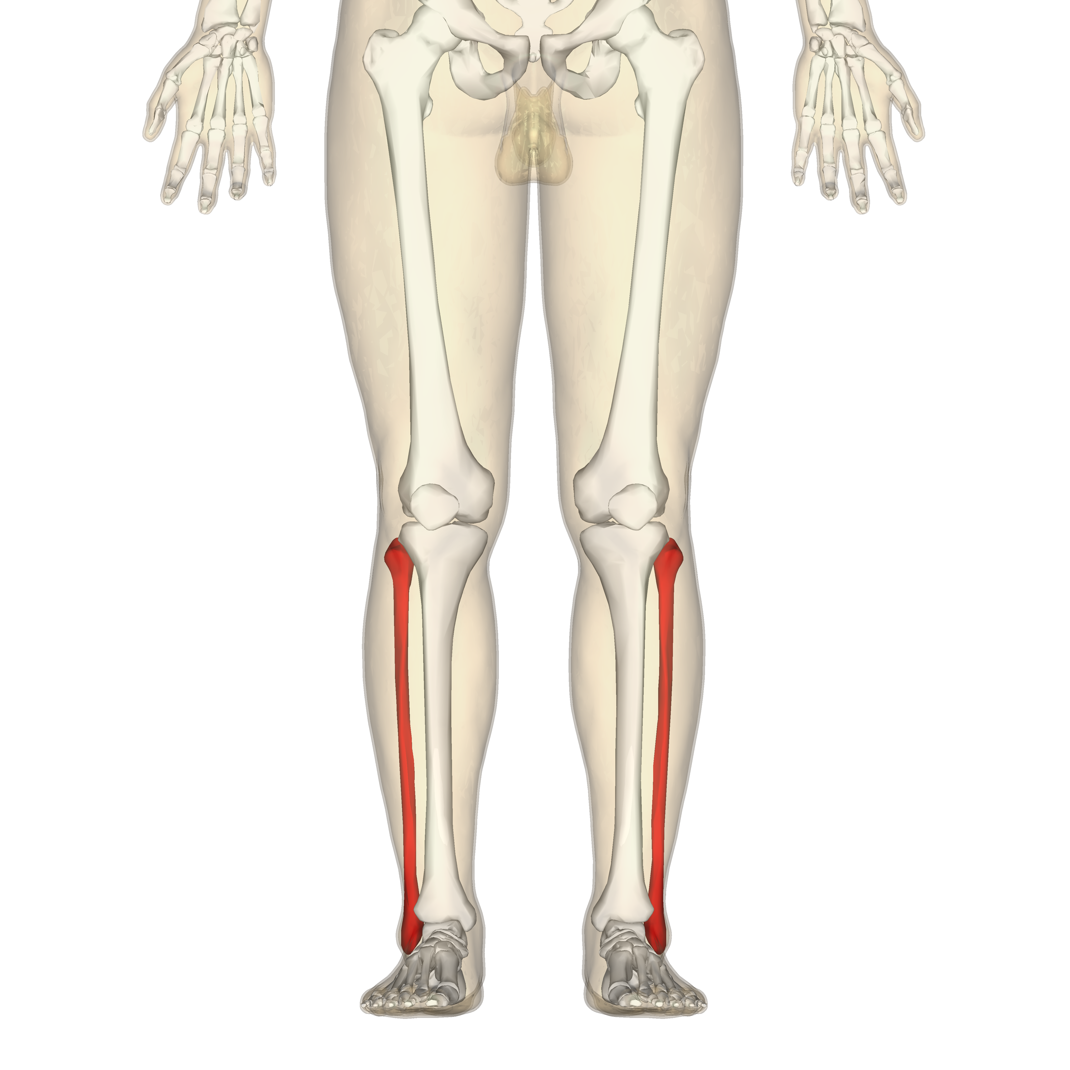
Fibula Wikipedia from upload.wikimedia.org When autocomplete results are available use up and down arrows to review and enter to select. These diagrams should explain and show you some of the basics. A horse's knee is several bones held together by small muscles, tendons, and ligaments. Beside that, we also come with more related ideas as follows free printable human anatomy coloring pages, lower leg muscle diagram blank and lower limb bones unlabeled. Neck length should be one third of the horse's total body length and equal the length of the horse's front leg. The coffin or pedal bone is the major hoof bone, supporting the majority of the weight. A human's knee joint is a hinge joint. Third phalanx or coffin bone is situated in the hoof wall leading on to the short pastern bone and aids the horse in supporting its weight.
Health diagram bone skeleton leg knee science anchor chart human human body.
The bones of the distal row are numbered, first to fourth carpal bones (66), of which the first is small and inconstant. It is to the pedal bone that the deep digital flexor tendon attaches. Neck length should be one third of the horse's total body length and equal the length of the horse's front leg. Beside that, we also come with more related ideas as follows free printable human anatomy coloring pages, lower leg muscle diagram blank and lower limb bones unlabeled. That way if you need to talk to a vet, or do a correct drawing, you'll have a solid foundation. The hoof of the horse contains over a dozen different structures, including bones, cartilage, tendons and tissues. The bones in the knee are similar to the bones of a human's wrists. See out article on horse vision for more info. Their leg bones are proportioned differently from those of a human. The pedal bone itself has an unusually high density of blood vessels within it. This page is mostly photos, graphs and charts about the horse. Similarly, the hock contains the bones equivalent to those in the human ankle and heel. Only mc2, 3, and 4 are present.
The horse leg anatomy in the rear includes the bones of the pelvis (the ilium, ischium and pubic bones), femur, tibia, fibula, metatarsus and the phalanxes leg bone diagram. Similarly, the hock contains the bones equivalent to those in the human ankle and heel.


0 Komentar